
The research team led by Aitziber L. Cortajarena has demonstrated that the new contrast agent for magnetic resonance imaging is safer for the heart than those normally used
The contrast agent is composed of minute clusters of iron atoms encapsulated in a custom-designed protein that is directed towards the affected area and blocks the processes that trigger the disease
San Sebastian, 29 October 2026. The Biomolecular Nanotechnology research group led by Ikerbasque research professor Aitziber L. Cortajarena has developed a new iron-based contrast agent for magnetic resonance imaging that can be used for both diagnosis and treatment. In a study carried out on lab-grown human heart tissue, it was proven that the contrast agent accumulates in the fibrotic tissue, that its signal intensity on the MRI image increases with the severity of the disease, and that it slows disease progression.
The study, led by Cortajarena and conducted in collaboration with Ana Villar’s group at the Institute of Biomedicine and Biotechnology of Cantabria (IBBTEC, UC-CSIC), has been published in the prestigious scientific journal Advanced Functional Materials. It paves the way for a possible alternative to gadolinium, the metal currently used in almost all contrast-enhanced MRI scans.
Cardiac fibrosis
Cardiac fibrosis is one of the main factors leading to heart failure, a disease that affects millions of people worldwide. There is currently no approved treatment specifically designed to slow down this condition. When the heart is damaged or under sustained strain (for example, after a heart attack or due to hypertension), a repair process begins, similar to the healing of a cut on the skin. The collagen production process is activated to repair and strengthen the tissue. However, this process often does not stop: the collagen continues to accumulate, the heart becomes stiff and finds it increasingly difficult to fill and pump the blood properly.
One of the techniques currently used for diagnosing fibrosis of the heart is MRI. In many cases, an intravenous contrast agent is administered to the patient, and is almost always based on gadolinium, which is retained in the damaged areas and makes them light up in the image. Although gadolinium works very well, it has various disadvantages. It is a metal that is foreign to the body. Its use is restricted in people with kidney failure, and it is known that small amounts of may be retained in different tissues for many years. This is especially concerning in patients who require repeated MRIs to monitor the progression of their disease, which is the case with cardiac fibrosis.
Iron atoms encapsulated in a custom-designed protein
For this reason, for many years researchers have sought to develop contrast agents based on iron, a metal that the body is familiar with and knows how to store and eliminate. However, there is a problem with existing iron-based contrast agents since, instead of illuminating the damaged area, they darken it, making it difficult to distinguish it in the images. Many of them have even been withdrawn from the market. The group led by Cortajarena, Scientific Director at CIC biomaGUNE, has been able to “overcome these problems by using designer proteins”, explains CIC biomaGUNE researcher Lydia Martínez Parra, lead author of the study.
Martínez adds, “the contrast agent consists of a specially designed protein that encapsulates and isolates minute clusters of iron atoms measuring approximately 1.4 nanometres (around 50,000 times thinner than a hair). It is also worth noting that this compound is safer for the body, is stable, releases very little iron and is of low toxicity to cells. Furthermore, it is easy and sustainable to manufacture, since it is produced in water, without toxic reagents or extreme conditions.”
A lab-grown fibrotic heart
The CIC biomaGUNE group has tested the efficacy of the contrast agent in cardiac tissue created with 3D tissue engineering using patients’ heart cells, developed by Ana Villar’s group (IBBTEC, UC-CSIC). These tissues replicate certain characteristics of cardiac fibrosis, such as the accumulation of collagen and increase in tissue stiffness, and enable new therapeutic strategies to be evaluated in more representative conditions than conventional cell cultures.
Thus, the research team has verified that the contrast agent moves towards and accumulates predominantly in the fibrotic tissue, that the signal on the MRI increases with the severity of the fibrosis, and that it slows its progression since it reduces the production and storage of collagen and prevents the tissue from becoming stiff. “That is, as well as indicating where the damage is, this material shows antifibrotic activity in the models studied”, says research professor Cortajarena. This is a ‘theranostic’ agent that offers diagnosis and treatment in the same material.”
Martínez points out that “this new compound enables the achievement of a contrast in magnetic resonance imaging comparable to that of gadolinium-based agents. It also retains this capacity in both magnetic fields commonly used in clinical practice and stronger magnetic fields used in preclinical research.” The compound formed by the iron atoms encapsulated by the protein make the tissue appear bright on the MRI.
The team behind the study affirms that “there’s still a way to go to reach the patient. For now, this new contrast agent has been tested in cells and human tissue in the laboratory. The next step will be to study it so that it can be used in clinical practice, to see how it is distributed and eliminated from the body and how it behaves on a real cardiac scanner. Our goal is that, in the future, patients with cardiac fibrosis, including those who cannot receive gadolinium, can have a safer imaging test that also forms part of their treatment.
Additional information
The project was funded by the State Research Agency (projects PID2024-156436OB-I00, PID2022-137977OB-I00, PID2025-175536OB-I00 and PDC2022-133345-I00, the latter with NextGenerationEU/PRTR funds), the European Research Council (ERC Proof of Concept NanoImaging-101069356) and IDIVAL (PREVAL 21/07).
Bibliographic reference
Lydia Martínez-Parra, Andrea García-Esnaola, Helena Soto, Gabriela Guedes, Lourdes Marcano, Diana H. Panchuelo, Irantzu Llarena, Ana V. Villar, Aitziber L. Cortajarena
Protein-Engineered Iron Nanoclusters as Hybrid Theranostic Materials for MRI Monitoring and Treatment of Cardiac Fibrosis
Advanced Functional Materials
DOI: 10.1002/adfm.78994
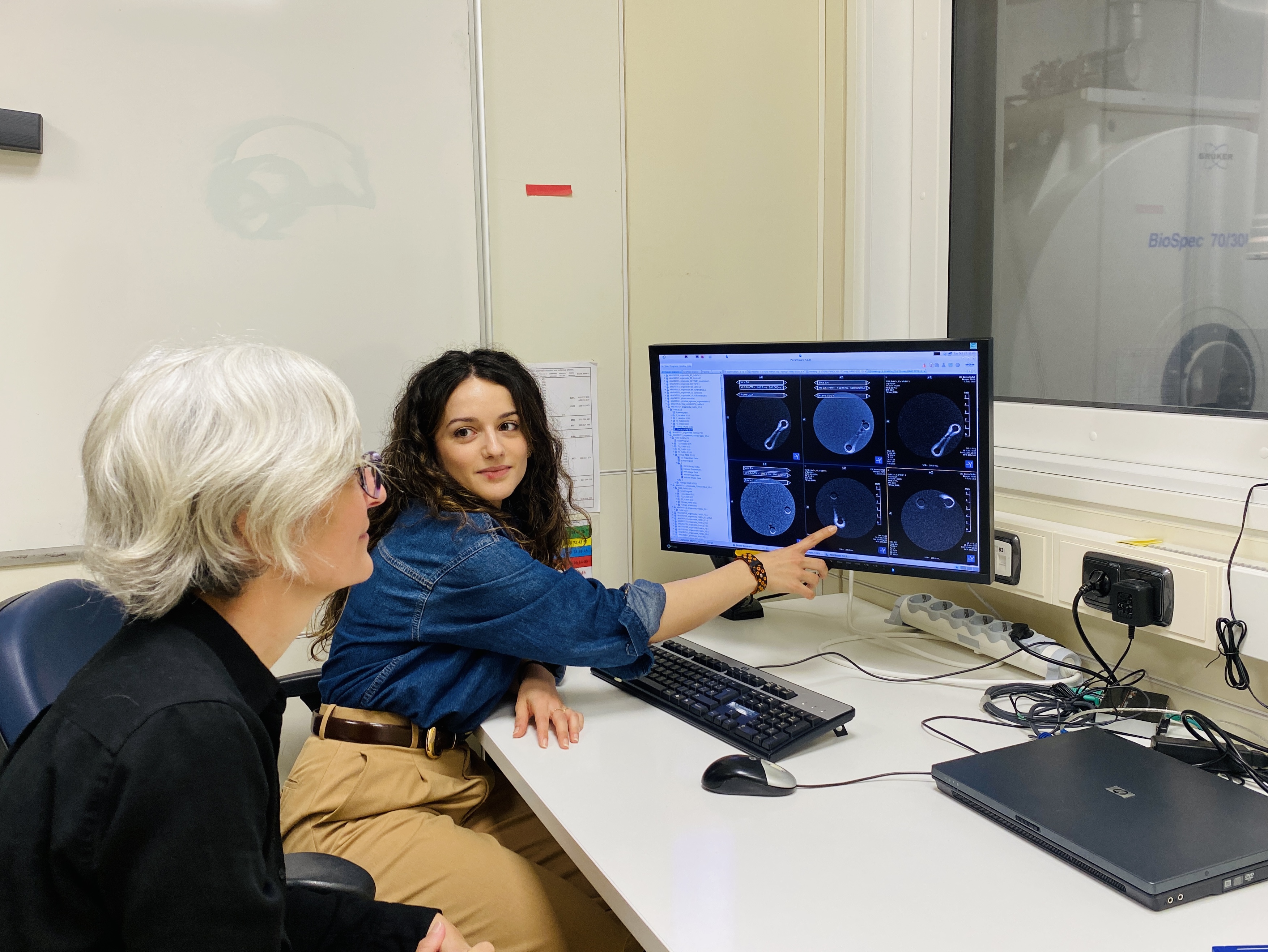
Photo captions
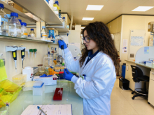
Photo 1. Postdoctoral researcher Lydia Martínez, from CIC biomaGUNE (Donostia/San Sebastián), prepares the compound to evaluate its properties as a magnetic resonance imaging (MRI) contrast agent.
Photo 2. Researchers Lydia Martínez and Aitziber L. Cortajarena, from CIC biomaGUNE (Donostia/San Sebastián), examine magnetic resonance images of three-dimensional models of human cardiac tissue acquired using a 7-tesla (7 T) preclinical MRI scanner.
About CIC biomaGUNE
The Centre for Cooperative Research in Biomaterials, CIC biomaGUNE, a member of the Basque Research and Technology Alliance (BRTA), is a non-profit research organisation in San Sebastian that carries out cutting-edge research at the interface between chemistry, biology and physics, with a particular focus on the study of the molecular-scale properties of biological nanostructures and their biomedical applications. In 2018, it was accredited as a María de Maeztu Unit of Excellence, for meeting the excellence criteria of having a significant impact and high level of competitiveness in its field of activity in the global scientific community.
Press Release
Nota de Prensa
Prentsa Oharra